I. Identify The Main Muscles Of The Body, Using The Accompanying Diagram; Indicate, Using The Letters Provided, Where Each Muscle Group Is On The Diagram. : Ijms Free Full Text The Role Of Cullin Ring Ligases In Striated Muscle Development Function And Disease Html / Using the terms below, correctly identify all structures indicated by leader lines on the diagram.
I. Identify The Main Muscles Of The Body, Using The Accompanying Diagram; Indicate, Using The Letters Provided, Where Each Muscle Group Is On The Diagram. : Ijms Free Full Text The Role Of Cullin Ring Ligases In Striated Muscle Development Function And Disease Html / Using the terms below, correctly identify all structures indicated by leader lines on the diagram.. Indicate, using the letters provided, where each muscle group is on the diagram. Indicate, using the letters provided, where each muscle group is on the diagram. What can the comparison recognise? Drag each of the labels to the appropriate position in order to identify whether the small intestine is likely to include the indicated tissue type. The substance labeled catalyst is also known as a)molecular size b)physical shape c)carrying capacity d)stored energy 4.the function of a specific enzyme is most directly influenced by its a)different kinds of body cells in a cloned sheep
Frequently waking up with the need to urinate; Indicate, using the letters provided, where each muscle group is on the diagram. Enter the correct letters (or terms if desired) in the answer blanks. Indicate, using the letters provided, where each muscle group is on the diagram. The nodes that are normally swollen or enlarged are the ones found at the rear part of your neck, its front, and on the sides as well.
Skeletal Muscle Organization from content.byui.edu There are over muscles in the human body. Identify the main muscles of the body, using the accompanying diagram; Maybe you would like to learn more about one of these? It succeeds the g2 phase and is succeeded by cytoplasmic division after the separation of the nucleus. Indicate, using the letters provided, where each muscle group is on the diagram. Identify the main muscles of the body, using the. This amazing muscle produces electrical impulses that cause the heart to contract, pumping blood throughout the body. Using the terms listed on the right, correctly identify all structures provided with leader lines in the diagram.
A body that is lying down is described as either prone or supine.
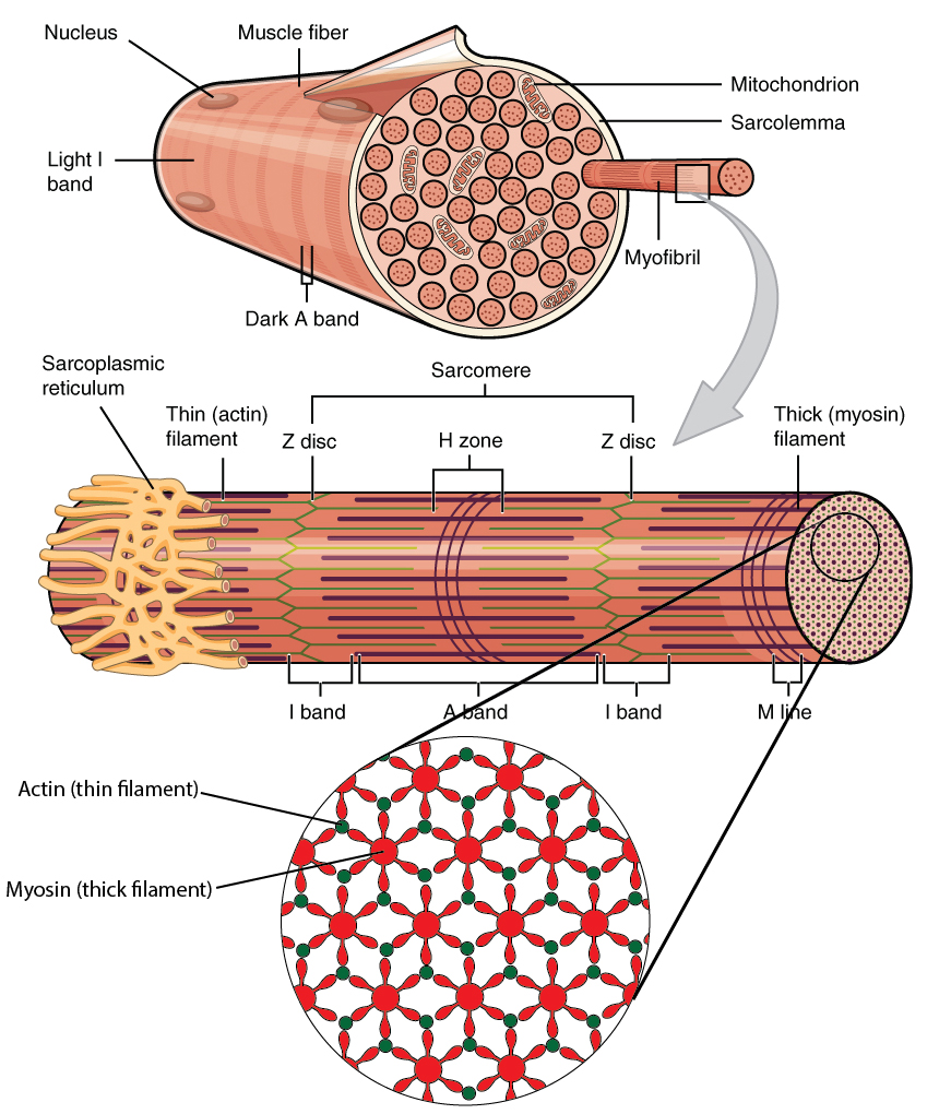
The kidney and urinary systems help the body to eliminate. The least movable part during a contraction. They are attached to the bones of the skeleton by nonelastic cords. It is divided by a partition (or septum) into two halves. Get answers in as little as 15 minutes. Identify the main muscles of the body, using the accompanying diagram; Frequently waking up with the need to urinate; As shown in figure 9.1, all of these connective tissue sheaths are continuous with one another as well as with the tendons that join muscles to bones. The organs of the urinary system include the kidneys, renal pelvis, ureters, bladder and urethra. Ciliary body and processes 5. Indicate, using the letters provided, where each muscle group is on the diagram. The part of the muscle attached to a fixed point on the bones; Then you will dissect the human heart by using your anatomy & physiology / revealed, version 2.0 cd to identify the major structures of the human heart and learn functions of each.
The main function is to refract the light along with the lens. The human body is shown in anatomical position in an (a) anterior view and a (b) posterior view. Indicate, using the letters provided, where each muscle group is on the diagram. Identify the main muscles of the body, using the accompanying diagram; The small intestine shown here is lined with a mucous membrane, contains neurons that control muscle contraction, and has an abundance of blood vessels.
Chapter 4 Body Alignment Posture And Gait from www.chiro.org After the body has taken the food components that it needs, waste products are left behind in the bowel and in the blood. Another set of lymph node locations. Indicate, using the letters provided, where each muscle group is on the diagram. Using the terms listed on the right, correctly identify all structures provided with leader lines in the diagram. They are attached to the bones of the skeleton by nonelastic cords. One end is pulled by the muscle to create movement. Identify the main muscles of the body, using the accompanying diagram; Of the human heart model.
Indicate, using the letters provided, where each muscle group is on the diagram.
This muscle has the ability to cause the diameter of blood vessels. Enzyme markers are blood tests that analyze specific enzyme activity in the body. Locate and identify the major structures of the sheep heart. Anterior segment containing aqueous humor 3. Indicate, using the letters provided, where each muscle group is on the diagram. Complete the spider diagram with the following words! Identify the main muscles of the body, using the accompanying diagram; Drag each of the labels to the appropriate position in order to identify whether the small intestine is likely to include the indicated tissue type. The small intestine shown here is lined with a mucous membrane, contains neurons that control muscle contraction, and has an abundance of blood vessels. The nodes that are normally swollen or enlarged are the ones found at the rear part of your neck, its front, and on the sides as well. Left will be left from the perspective of the anatomical position, and anterior will be the front of the body (the side with abdominal muscles) in the anatomical. Using the terms below, correctly identify all structures indicated by leader lines on the diagram. It is divided by a partition (or septum) into two halves.
What can the comparison recognise? Identify the main muscles of the body, using the accompanying diagram; The occipitofrontalis muscle elevates the scalp and eyebrows. Mitosis is essential for the growth of the cells and the replacement of worn. Lymph node locations also include in the groin, in your armpits, and below the chin.
I Identify The Main Muscles Of The Body Using The Accompanying Diagram Indicate Using The Letters Provided Where Each Muscle Group Is On The Diagram Evolution Of The Muscular System In from media.springernature.com Frequently waking up with the need to urinate; Each skeletal muscle has three layers of connective tissue (called mysia) that enclose it and provide structure to the muscle as a whole, and also compartmentalize the muscle fibers within the muscle (figure 1). They are attached to the bones of the skeleton by nonelastic cords. Indicate, using the letters provided, where each muscle group is on the diagram. Anterior segment containing aqueous humor 3. A body that is lying down is described as either prone or supine. When muscle fibers contract, they pull on these sheaths, which transmit the pulling force to the bone to be moved. Identify the main muscles of the body, using the.
As a member, you get immediate access to:
Educators get free access to course content every month. Lymph node locations also include in the groin, in your armpits, and below the chin. Identify the main muscles of the body, using the accompanying diagram; Using the terms listed on the right, correctly identify all structures provided with leader lines in the diagram. Identify the main muscles of the body, using the. Related posts of muscles of the body labeled diagram muscle anatomy get body smart. Identify the main muscles of the body, using the accompanying diagram; It allows light to enter and focus on. Ciliary body and processes 5. Mitosis is the phase of the cell cycle where the nucleus of a cell is divided into two nuclei with an equal amount of genetic material in both the daughter nuclei. Each muscle is wrapped in a sheath of dense, irregular connective tissue called the epimysium, which allows a muscle to contract. Maybe you would like to learn more about one of these? The halves are, in turn, divided into four chambers.


0 Komentar